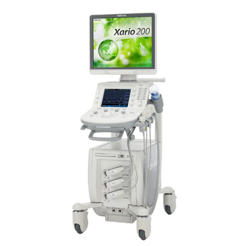
Toshiba’s Xario 200 ultrasound system takes the worry out of providing high-quality patient care with a smaller, compact ultrasound system. Combining advanced imaging technologies with industry-leading depth and detail, the Xario 200 delivers outstanding clinical performance for routine and advanced studies in the widest range of clinical applications. The system includes an array of ergonomic enhancements automated features to enhance your productivity.
The Xario 200 provides exceptional cost of ownership with the industry’s best technology protection program. All with a sleek, compact system that’s fully loaded and ready to roll into almost any space.
Xario 200’s High-Density Beamformer Architecture produces advanced digital signal processing technology to reveal clinical information never seen before. With comprehensive image enhancement capabilities and an unsurpassed 40 cm depth setting, Toshiba is clearly the leader in delivering best-in-class images for all patient exams.
- Precision Imaging enhances the definition and sharpens the edges of
structures to separate clinical information from noise.
Differential Tissue Harmonic imaging (D-THI)*
- D-THI increases contrast and spatial resolution at greater depths and on difficult-to-image patients.
ApliPure+
- ApliPure+ achieves unparalleled uniformity and detail while preserving clinically significant markers.
Advanced Dynamic Flow (ADF)*
- ADF displays smallest blood vessels and complex blood flow with unequaled accuracy and detail.
Xario 200 systems also feature advanced imaging technologies and quantification tools that extend diagnostic capabilities, increasing confidence in your clinical decision making.
Volume Imaging Suite*
- Volume Imaging Suite captures volume data sets at high—volume rates for shorter exam times and features a comprehensive set of imaging modes (Surface rendering, Muitiview and MPR).
Stress Echo
- Stress Echo enables fast and accurate wall motion assessment and supports both standard and user-definable protocols for exercise and pharmacological stress studies.
Auto- IMT
- Auto-IMT provides an easy-to-use automation tool for measuring intima-media thickness (IMT) of the proximal and distal arterial walls to help determine a patients risk for cardiovascular disease.
Real-time Elastography*
- Real-time Elastography provides a visual representation (color mapping) of the elasticity of lesions following manual compression and helps localize and assess palpable masses with exceptional accuracy, sensitivity and reproducibility.